Coptis chinensis rhizomes (CR) are one important ingredient of traditional Chinese herbal formulas such as San-Huang-Xie-Xin-Tang which is used for treatment of cardiovascular and neurodegenerative diseases. Recent studies suggest that the extract of CR might be a potential therapeutic agent for amelioration of neurological disorders associated with oxidative stress. In the present study we aimed at revealing the main active compound(s) of the CR extract and at investigating the mechanism of action. Four main alkaloids of the CR extract (berberine, coptisine, jatrorrhizine, and palmatine) were selected for this study. Results showed that out of those alkaloids only pretreatment with coptisine significantly attenuated tert-butylhydroperoxide induced reduction of cell viability, increased rate of apoptosis, and declined mitochondrial membrane potential. Elisa assay and quantitative real-time PCR analyses revealed that thioredoxin-interacting protein (TXNIP) gene expression was downregulated by coptisine, which could explain the neuroprotective effect, hypothetically, by strengthening the thioredoxin defense system against oxidative stress and attenuation of apoptosis signal-regulating kinase (Ask1) mediated apoptotic signaling. A comparison between coptisine and CR extract identified coptisine as the main single component responsible for the neuroprotective effect. Based on the results the CR extract and coptisine are promising candidate agents for prevention or improvement of diabetic neuropathy and neurodegenerative disorders.
Coptis chinensis (Franch) rhizomes (CR), commonly known as Coptidis rhizoma or “Huang Lian,” has been used in Traditional Chinese Medicine (TCM) since ancient times and was recommended by famous physicians in TCM history like Li Shizhen, Tang Shenwei, and Tao Hongjing for inflammatory diseases.
CR is a main ingredient of multiple historical prescriptions, for example, San-Huang-xie-xin-tang (SHXT), which originated in the Ming dynasty. SHXT is composed of Coptis chinensis (Franch) (Coptidis rhizoma),Rheum officinale Baill (Rhei rhizoma), and Scutellaria baicalensis Georgi (Scutellariae radix). Recent studies showed that SHXT has neuroprotective properties due to its anti-inflammatory and antioxidative effects. It has been shown that one of the main ingredients of SHXT, the dried root CR also known as Chinese goldthread (huang-lian in Chinese), is effective for the treatment of neurodegenerative disease associated with oxidative stress. Some of its single compounds showed neuroprotective, neuroregenerative, anti-apoptotic, and antioxidative effects which strongly point out that CR is one main component in decoctions used for the treatment of oxidative stress associated with neurodegenerative disease.
Reactive oxygen species (ROS) have a significant impact on the development of neurodegenerative disease like Alzheimer or Parkinson's disease. Cells including neurons are usually well-protected from ROS-induced cytotoxicity by the endogenous antioxidant system. However, if the oxidative stress exceeds the antioxidative capacity of this system it can lead to deoxyribonucleic acid (DNA) demethylation, histone acetylation, oxidative protein and lipid modification, increase of intracellular calcium ions (Ca 2+), depolarization of the mitochondrial membrane potential (MMP), and release of cytochrome C into the cytosol and, as a consequence, to apoptosis or necrosis. For this reason, therapeutic strategies targeting ROS-induced cytotoxicity are needed and could have a major impact on the treatment of neurodegenerative diseases, which are associated with oxidative stress.
We recently showed that 2 h and 24 h pretreatment of SH-SY5Y neuroblastoma cells with the watery extract of CR (CRE) significantly attenuated tert-butylhydroperoxide- (t-BOOH-) induced cytotoxicity. Further statistical analysis revealed that 24 h pretreatment with CRE was more effective than 2 h pretreatment.
The present study aimed at revealing the main active compound of CRE, which is responsible for the cytoprotective effect of the extract, and at comparing the effectiveness of the single compounds with the whole extract.
tert-Butylhydroperoxide (t-BOOH), thiazolyl blue tetrazolium bromide (MTT), and dimethyl sulfoxide (DMSO) were purchased from Sigma (Taufkirchen, Germany). 2′,7′-Dichlorodihydrofluorescein diacetate (H 2 DCFH-DA), Mitotracker Red CMX Ros, and Hoechst 33342 were obtained from Life Technologies (Darmstadt, Germany). Berberine (Ber), coptisine (Cop), jatrorrhizine (Jat), and palmatine (Pal) were ordered from Cfm Oskar Tropitzsch (Markdredwitz, Germany). All other reagents were purchased from Roth (Karlsruhe, Germany).
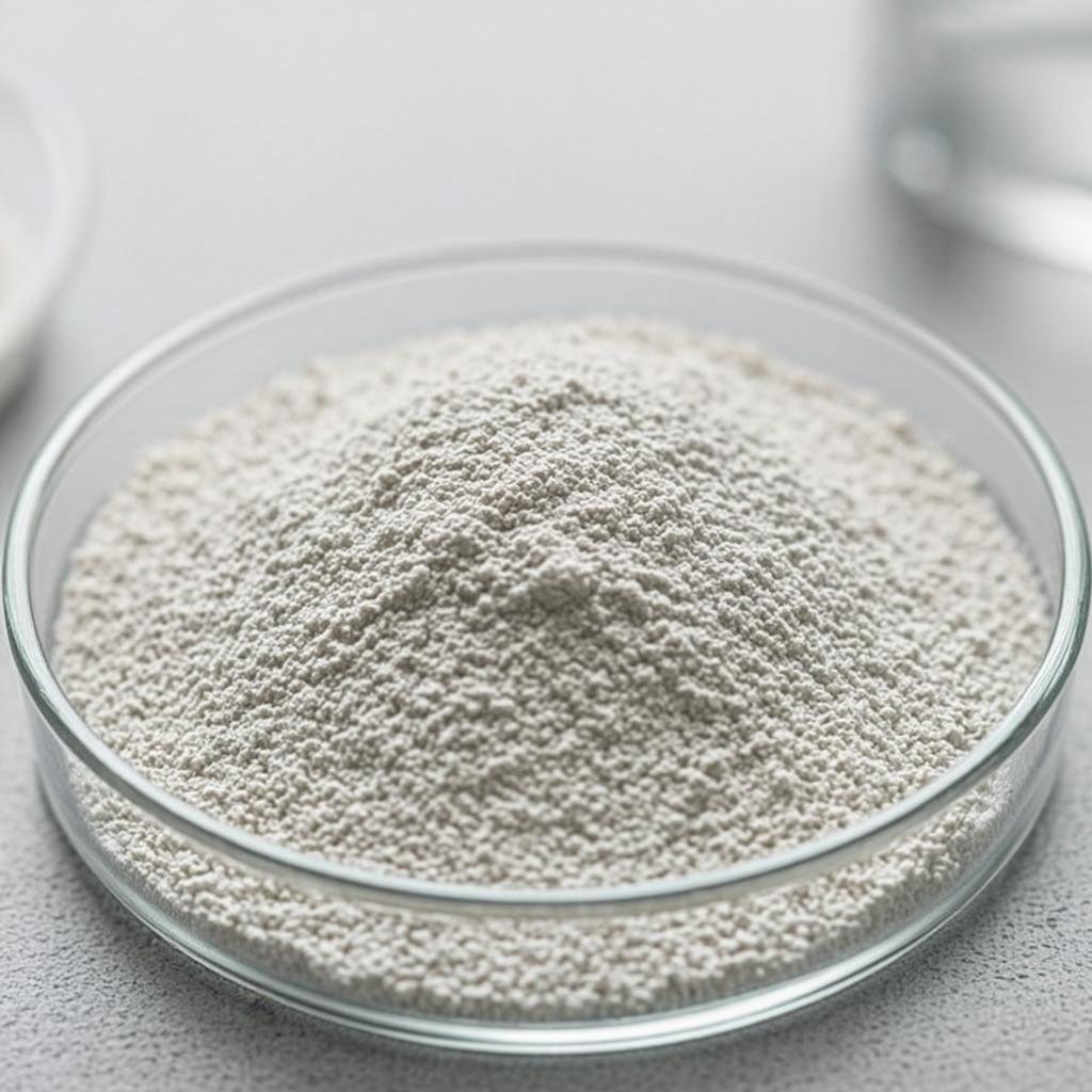
Coptis chinensis(Franch) was obtained from China Medica (Ch. B. 930034; 83684 Tegernsee, Germany), as dried rhizome. Identity and purity were confirmed according to the Pharmacopoeia of the People's Republic of China. Sebastian Kneipp research laboratory for residue analysis and organic trace analysis (Bad Wörishofen, Germany) certified that heavy metal, pesticide, and microbiological contamination were below the guideline of the Pharmacopoeia Europaea and Regulation (EC) Number 396/2005 of the European Commission.
Coptis chinensis extract was prepared as described previously. 10 g of grounded rhizome was boiled in 100 mL distilled deionized water (DDW) for 30 minutes and the extract was centrifuged afterwards. Supernatant was collected and the residue was extracted a second time with 100 mL DDW. Combined supernatants were dried with a rotary-vacuum evaporator (60°C, 200 mbar; Rotavapor-R, Büchi) and a vacuum concentrator (Bachofer). Dried extracts were stored at −20°C until use.
High-performance liquid chromatography (HPLC) analysis was performed according to the method described previously. Briefly, chromatographic separation was conducted on an Alltima C18 (250 mm × 4.6 mm × 5 μ m, S/N: 213100139, temperature: 25°C) column with 0.1% trifluoroacetic acid (A) and acetonitrile (B) as mobile phase and at a flow rate of 1 mL/min. Berberine (Ber), coptisine (Cop), jatrorrhizine (Jat), and palmatine (Pal) were used as reference standard compounds.
Human neuroblastoma SH-SY5Y cells were cultivated in RPMI 1640 medium containing 10% fetal calf serum, 100 U/mL penicillin, and 100 μ g/mL streptomycin. Cells were grown in a humid atmosphere of 5% CO 2 and 95% air at 37°C. All cell culture reagents were obtained from Sigma (Taufkirchen, Germany).
Cell viability was determined by the MTT assay. Ber, Cop, Jat, and Pal stock solutions were diluted in medium to their final concentration and sterilely filtrated, and different concentrations were added at the start of the incubation time for 24 h. Afterwards, cells were incubated for 2 h with 100 μ M t-BOOH. Medium containing t-BOOH was removed, cells were washed with Dulbecco's phosphate-buffered saline (DPBS), and 1 mM MTT solution was added for 2 h. Subsequently, 100 μ L 2-Propanol was added and the plate was agitated for 1 h at 450 revolutions per minute (rpm) at room temperature (RT). Absorption was measured 3 times at 570 nm.
Reactive oxygen species in the cells were measured with the 2′,7′-dichlorodihydrofluorescein diacetate probe (H 2 DCFH-DA). SH-SY5Y cells were seeded into a 96-well microplate (4 ∗ 10 4 cells/well) and incubated with 100 μ g/mL CRE or 20 μ M Cop for 24 hours. Afterwards, fresh medium containing 20 μ M H 2 DCFH-DA was added for 30 min at 37°C in the dark. Subsequently, cells were washed with DPBS and medium containing 100 μ M t-BOOH was added. Fluorescence was measured every 10 minutes for 120 minutes (excitation: 485 nm; emission: 528 nm).
Apoptotic nuclei and mitochondrial membrane potential were measured using Mitotracker Red CMX Ros and Hoechst 33342. Cells were stained first with 25 nM Mitotracker Red CMX Ros for 1 hour, followed by 20 minutes fixation with 4% paraformaldehyde (PFA) and 10 minutes Hoechst 33342 (4 μ M) staining. Images were captured with a Leica microscope and an Axiovision camera. To determine MMP, the intensity sum of the Mitotracker Red CMX Ros fluorescence was measured for each cell. Apoptosis was detected by analyzing the morphology of the Hoechst 33342 stained nuclei. Experiments were repeated 3 times and at least 1600 cells were analyzed for each group.
Total RNA was isolated with the RNeasy MINI Kit (Quiagen; Hilden; Germany) according to the manufacturer's instructions. Reverse transcription was carried out with the high capacity RNA-to-cDNA Kit (Applied Biosystems). For semiquantitative analysis, LightCycler 480 SYBR Green 1 Master (Roche) and the following human specific primers were used: TXNIP 5′-GATCACCGATTGGAGAGCCC-3′ and 5′-TGCAGGGATCCACCTCAGTA-3′; GAPDH 5′-GCATCTTCTTTTGCGTCGCC-3′ and 5′-CCCAATACGACCAAATCCGTTG-3′. Obtained data were analyzed as previously described.
Cells were seeded in T-25 flasks (2.5 × 10 6 cells/flask) and treated for 24 hours with CRE or Cop. Total proteins were isolated with mammalian cell lysis reagent (CellLytic M) according to the manufacturer's instructions and stored at −80°C until use.
Protein concentration was determined with Roti-Quant following the protocol provided by the manufacturer. Human TXNIP protein concentration was measured with the CircuLex Human TXNIP ELISA Kit (MBL international cooperation, Biozol, Germany) following the instructions of the provided protocol. Experiments were repeated three times and each sample was measured in duplicates.
Data are presented as means ± standard error of the mean (SEM) of n experiments. Statistical significance between groups was determined with OriginLab pro 8.5 by ANOVA, followed by the Bonferroni Post Hoc Test. P< 0.05 was considered as statistically significant.
Watery extraction of CR yields 21.5% by weight of the dried herb. The CRE was composed of 554.9 ± 14.6 mg/g Ber, 60.6 ± 0.5 mg/g Cop, and 51.9 ± 0.2 mg/g Pal. Representative high-performance liquid chromatogram of the extract and of the mixed standard compounds is shown.
To investigate the neuroprotective effect of CR main alkaloids against t-BOOH-induced toxicity, we first examined if those single components exhibited any cytotoxic effect in the concentration range between 0.1 and 40 μ M. Results showed no significant cytotoxic effect of Ber, Cop, Jat, and Pal in comparison to the medium control (P< 0.01). Treatment of cells with 100 μ M t-BOOH resulted in significant decrease of cell viability to 53.2 ± 1.7% (P< 0.01; versus medium control). Pretreatment of the cells with Ber, Jat, and Pal (0.1–40 μ M) before t-BOOH-induced oxidative stress showed no significant protective effect compared to the t-BOOH control. However, pretreatment with 1–40 μ M Cop for 24 hours resulted in a significant increase of cell viability from 53.2 ± 1.7% (t-BOOH control) up to 65.6 ± 2.6% (40 μ M).
Previously, we showed that 24-hour pretreatment with the watery extract of CR had a significant protective effect