Testosterone (TS) is an important androgen drug and a precursor of steroid drug synthesis. Ketoreductase 2 (KR-2) (GenBank accession no. ABP64403.1) is observed to stereo-selectively catalyze the bioreduction of 4-androstene-3,17-dione (4-AD) to testosterone and contribute to the regeneration of NADH using isopropanol as a co-substrate. The Km value of KR-2 was 2.22 mmol/L with 4-AD, and the optimal pH was 6.5–7.0. The enzyme is stable and demonstrates relatively high-level enzyme activity at 40 °C. Acetone significantly inhibits this activity. This inhibition was overcome using an intermittent vacuum during the reaction process. Finally, the amount of TS reached 65.42 g/L after a 52 h reaction with 65.8 g/L 4-AD, 10% isopropanol, and 2 g/L β–NAD+ at 40 °C, with a conversion rate of 98.73%. A total of 6.15 g of TS was obtained from 6.58 g of 4-AD after the reaction and purification; the HPLC purity was 99.82%, and the overall yield was 92.81%. This enzyme provides a promising route for the green biosynthesis of testosterone for industrial applications.
Testosterone (TS) is an androgen drug with important physiological activity [1,2] and a key intermediate for synthesizing many high-value steroid drugs, such as methyltestosterone, testosterone heptate, and 6β-hydroxytestosterone [3].
In previous studies, testosterone has been chemically synthesized from indanone [4], olefinic nitrile oxide [5], and androstene-3,17-dione [6]. In the newer reports, the synthesis of testosterone was mostly carried out using starting intermediates with a cyclopentane–polyhydrophenanthrene structure, such as cholesterol, diosgenin, phytosterols, 4-dienosterone-3,17-dione (4-AD), etc. These intermediates can be biologically transformed into testosterone, and the process is relatively simple and cost-effective [7,8,9,10]. When 4-AD was used as the starting intermediate, testosterone was obtained after one-step enzymatic reduction, which is the preferred method for the industrial production of TS.
In mammals, TS is synthesized from 4-AD catalyzed by 17β-hydroxysteroid dehydrogenase (17β-HSD) [11]. In 2016, Shao et al. [12] co-expressed human 17β-HSD and Saccharomyces cerevisiae glucose-6-phosphate dehydrogenase (G6PDH) in Pichia pastoris GS115, and the co-expressed system produced 11.6 g/L of TS from 4-AD in 120 h. In 2021, Ding et al. [13] improved the activity of 17β-HSD using a rational design, and the TS yield increased by 197% to 3.98 g/L.
The 17β-HSD from fungi and bacteria has been reported for the enzymatic reduction in 4-AD to TS. In 2017, Fernandez-Cabezon et al. [14,15] cloned the genes encoding 17β-HSDs from the bacterium Comamonas testosteroni and the fungus Cochliobolus lunatus, and the engineered strains of Mycobacterium smegmatis produced high yields of TS from sterols or androst-4-ene-3,17-dione (AD). In 2019, Govinda Guevara et al. [16] cloned 17β-HSD from Cochliobolus lunatus into Rhodococcus ruber Chol-4, and TS was synthesized from 4-AD, with a molar conversion rate of 61% using glucose for co-factor regeneration. The 4-AD can be synthesized from phytosterols. In 2022, DN. Tekucheva et al. [17] reported the two-stage transformation of phytosterol by the actinobacteria Mycolicibacterium neoaurum VKM Ac-1815D and Nocardioides simplex VKM Ac-2033D, which were capable of oxidizing the phytosterol side chain and reducing androstenedione at C17, respectively. A total testosterone yield of 53% was obtained using 10 g/L of phytosterol.
The 17β-HSD belongs to the short-chain dehydrogenase (SDR) family [18,19]. In addition to 17β-HSD, a few other SDRs have been reported for the biotransformation of 4-AD to testosterone. Zhou et al. [20] reported 4-AD transformation via alcohol dehydrogenase from Ralstonia sp. (RasADH) and commercial glucose dehydrogenase (GDH). A TS space–time yield of 1.65 g/L/h was achieved at a load of 10 g/L 4-AD. In 2022, Su et al. [21] reported that the Prelog enzyme from Pseudomonas can reduce 4-AD to TS in diastereomeric excess. They co-expressed this Prelog enzyme and formate dehydrogenase in Escherichia coli BL21 (DE3), resulting in a testosterone yield of 28.8 g/L.
These studies utilized formate dehydrogenase (FDH) and GDH to regenerate the co-enzyme NADH. No reports on the use of secondary alcohol dehydrogenase for co-enzyme regeneration in TS biosynthesis have been published. During the biosynthesis of dehydroepiandrosterone (DHEA) using 5-androstene-3,17-dione (5-AD), we unexpectedly discovered a secondary alcohol reductase that can reduce the 17 carbonyl groups of 5-AD to hydroxyl groups, and TS was found to be an impurity in the reaction mixture (Figure 1a). This secondary alcohol dehydrogenase (KR-2) belongs to ketoreductase and was formerly used as a bifunctional enzyme in the synthesis of the antiviral drug atazanavir [22] (Figure 1b). It catalyzed the reduction in the intermediate (3S)-1-chloro-2-oxo-3-(N-tert-butyloxycarbonyl)-4-phenylbutane (α-chloroketone) to (2R,3S)-1-chloro-2-hydroxy-3-(N-tert-butyloxycarbonyl)-4-phenylbutane (α-chlorohydrin) and also performed NADH regeneration by dehydrogenating isopropanol to acetone.
Here, we performed TS biosynthesis from 4-AD with ketoreductase. It is interesting that TS was formed as the sole product when only ketoreductase 2 (KR-2) was used in the reaction, with 4-AD as the starting compound (Figure 1c). In this study, the TS concentration reached 65.42 g/L from 4-AD using KR-2, which is currently the highest reported level.
The 4-AD was purchased from Goto Biopharm (Shiyan, China). The β–NAD+ oxidized form (free acid, NAD+) was from Sangon Biotech Co., Ltd., Shanghai, China. KH 2 PO 4 and K 2 HPO 4 were purchased from Aladdin Reagent Co., Ltd. (Shanghai, China). Tryptone and yeast extracts were purchased from Angel Yeast Co., Ltd. (Yichang, China). All the other reagents used in this study were of analytical grade.
The GenBank accession no. for KR-2 was ABP64403.1. The recombinant bacterium Escherichia coli BL21 (DE3)/pET-TZU5 cells were constructed and preserved at the Enzyme Engineering Laboratory of Taizhou University (Taizhou, China). Fermentation was performed in a 5 L bioreactor (Biotech-3BG, Shanghai BaoXing Bio-Engineering Equipment Co., Ltd., Shanghai, China). Low-temperature induction with α-lactose was adopted to achieve a high protein expression level [23,24]. The E. coli BL21(DE3)/pTZU-5 strain was grown overnight at 37 °C at 220 rpm in 5 mL Luria broth (LB) with 100 μg/mL ampicillin. The overnight meat soup was inoculated in 100 mL of TB (2.4% yeast extract, 1.2% tryptone, 0.4% glycerol, 17 mmol/L KH 2 PO 4, and 72 mmol/L K 2 HPO 4) with 100 μg/mL ampicillin in disposable baffled flasks and grown at 37 °C at 220 rpm for approximately 4–5 h until the cells reached OD 600 0.6–0.8. This preculture was used to seed 3.5 L TB medium in a 5 L fermenter. The bioreactor was run at a controlled temperature of 37 °C with >25% dissolved oxygen, and the aeration rate was maintained at 3.0 vvm. The culture was performed until the cell population reached OD 600. Subsequently, the culture temperature was reduced to 25 °C, and 150 mL of 20% (w/v) α-lactose solution was added. Agitation cascade was employed to maintain the dissolved oxygen (DO) at >25%. Approximately 15 h of fermentation was required until the cell population reached OD 600 stability.
For the KR-2 activity assay, a 25 μL crude enzyme sample was mixed with a 0.975 mL reaction substrate (pH 7.0 50 mmol/L KH 2 PO 4, 50 mmol/L K 2 HPO 4, 0.1 mL isopropanol, 2 g/L 4-AD, and 0.5 g/L NAD+) at 40 °C via shaking at 1100 rpm for 20 min in a metal bath. The sample was diluted 2–5-fold with a diluent (acetonitrile–water containing 0.1% phosphoric acid), centrifuged at 10,000× g for 2 min, and analyzed using HPLC.
The activity of KR-2 was determined based on testosterone production. One unit of KR-2 is defined as the amount of enzyme required to catalyze the production of 1 μmol of testosterone per minute at 40 °C. The assay result was linear when the specific activity level of the sample was lower than 6.0 U/mL. The samples were diluted to ensure the result was lower than 6.0 U/mL after dilution.
The optimization of the reaction conditions was performed in a 50 mL centrifuge tube; 2.0 mL of enzyme solution was mixed with 8 mL of reaction substrate mixture (containing 10 g/L of 4-AD, 1 mL of isopropanol, 1 g/L of NAD+, and 7 mL of 50 mM pH 7.0 potassium phosphate buffer). The reaction was performed at 40 °C and 200 rpm in a water bath. The samples were diluted 10–100-fold and centrifuged at 10,000 rpm for 2 min for HPLC analysis.
A scaled-up reaction was conducted in a 250 mL four-necked flask. The reaction mixture contained 1.0–6.58 g of 4-AD, 10 mL of isopropanol, 100–200 mg of NAD+, 20 mL of crude enzyme, and 70 mL of 50 mM pH 7.0 potassium phosphate buffer, with a total volume of 100 mL. The reaction mixture was adjusted to pH 7.0 with 1 mol/L NaOH. The reaction was performed at 40 °C in a water bath.
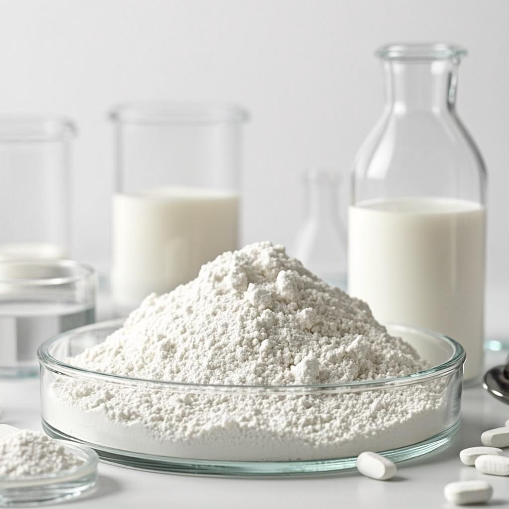
To isolate and extract the testosterone, 300 mL of ethanol was added to the reaction solution. The mixture was then filtered, and the filtrate was concentrated under a vacuum to remove the solvent. The precipitate was recovered via filtration and recrystallized in ethanol. White powder was obtained and dried overnight at 50 °C to obtain the final product.
The HPLC analysis of testosterone and 4-AD was performed as described by Han, with certain modifications [25]. The HPLC system used was the Agilent 1260 Infinity II HPLC system (Agilent Ltd., Santa Clara, CA, USA). A C18 (Supersil ODS2 5 μm 250 × 4.6 mm) reversed-phase analytical column was used at 50 °C. The mobile phase comprised 0.1% phosphoric acid and acetonitrile (40:60 v/v). The flow rate was 1 mL/min, and the detection wavelength was 240 nm. The retention times for testosterone and 4-AD were 5.697 min and 6.790 min, respectively.
To determine the optical rotation, the product (0.1 g) or testosterone standard was dissolved in 25 mL of ethanol, and the volume was increased to 100 mL with ethanol. The solution was loaded into a 20 cm long test tube, and optical rotation α was determined at 20 °C (589.44 nm). Nuclear magnetic resonance (NMR) analysis was performed at 101 MHz using chloroform [26].
The fermentation of engineered E. coli BL21 (DE3)/pET-TZU-5 in a 5 L fermenter occurred similarly to a previously published procedure [27,28]. The enzyme activity is tightly coupled with cell growth (Figure 2a); both increased quickly at approximately 3–8 h and then slowly from 8 to 13 h. At the end of fermentation (13 h), the enzyme activity level was 1121 U/L, and the target protein accounted for approximately 24.5% of the total protein content. SDS-PAGE analysis showed that approximately 74% of the target protein was in the supernatant (Figure 2b).
KR-2 was very efficient in catalyzing α-chloroketone to α-chlorohydrin [22], but 4-AD has not been used as a substrate for KR-2 yet. Compared to α-chloroketone, the pH range of the enzyme became more acidic when the substrate was 4-AD. The optimal pH was 6.5–7.0, and approximately 70% of enzyme activity was retained at a pH range of 6.0–9.0 (Figure 3a).