This article discusses the measurement of protease activity using a standardized assay. The assay involves the digestion of casein by proteases, resulting in the release of tyrosine, which can be quantified.
Tyrosine is liberated during the digestion of casein by proteases and serves as a measurable indicator of protease activity.
Absorbance is measured using a spectrophotometer after the reaction of tyrosine with the colorimetric reagent.
Key reagents include casein, trichloroacetic acid, and the colorimetric reagent for tyrosine.
The standard curve correlates known concentrations of tyrosine with absorbance values to quantify protease activity.
Yes, the assay is designed to be universally applicable for various proteases.
Proteases break peptide bonds. In the lab, it is often necessary to measure and/or compare the activity of proteases. Sigma's non-specific protease activity assay may be used as a standardized procedure to determine the activity of proteases.
Proteases break peptide bonds in the lab. It is often necessary to measure and or compare the activity of proteases Sigma's. Universal protease activity assay may be used as a standardized procedure to determine the activity of proteases, which is what we do during our quality control procedures.
In this assay, Casian acts as a substrate when the protease we are testing digests casian. The amino acid tyrosine is liberated along with other amino acids and peptide fragments. Free tyrosine then reacts with fallen and cyl cals phenol or fallen reagent to produce a blue colored chromophore, which is quantifiable and measured as an absorbence value on the spectrophotometer.
The more tyrosine that is released from casian, the more the chromophores are generated and the stronger the activity of the protease Absorbence values generated by the activity of the protease are compared to a standard curve which is generated by reacting known quantities of tyrosine with the FC reagent to correlate changes in absorbance. With the amount of tyrosine in micromoles from the standard curve, the activity of protease samples can be determined in terms of units, which is the amount of micromoles of tyrosine equivalence released from Cain per minute. Hello, my name is Carrie Cup Vineyard and I'm a scientist at our Sigma Aldridge DeKalb facility here in St.Louis, Missouri.
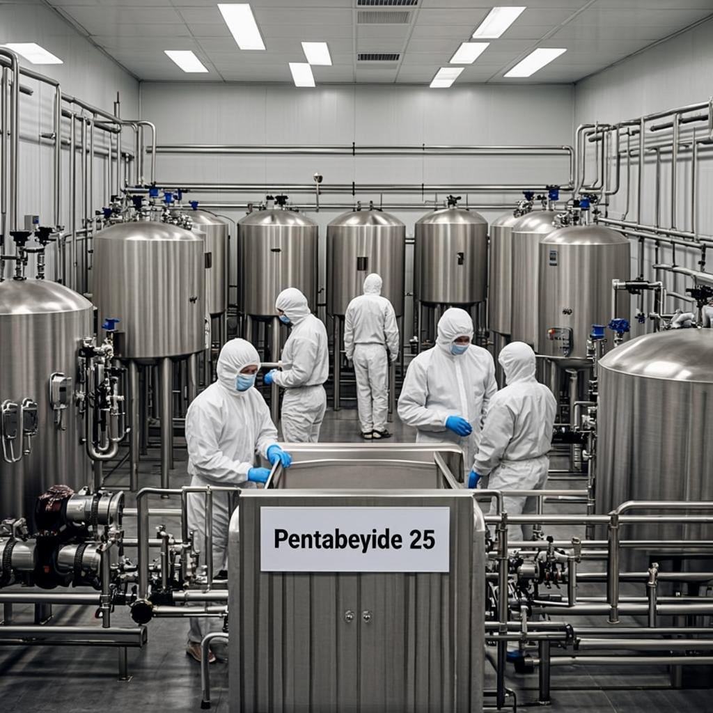
Today, I will show you a universally applicable way to test protease activity at Sigma Aldridge. We mainly use this assay in our quality control area to ensure that our protease has sufficient activity before we ship it directly to you at your lab. Before beginning the assay, we need to make sure that the following reagents are correctly prepared.
Now that we've prepared our reagents and they're all at the correct temperature, we will begin our protease assay.
To begin this assay, find suitable vials that will hold about 15 milliliters. For each enzyme that you will test, you will need four vials. One vial will be used as a blank, and three others will be used to assay Activity of three.
Dilution of the protease three dilution are useful when checking our final calculations against each other to each set of four vials at five milliliters of our 0.65%kasian solution and let them equilibrate in a water bath 37 degrees Celsius for about five minutes. Then add varying concentrations of our enzyme solution to three of the test sample vials, but not the blank. Mix them by swirling and incubate for 37 degrees Celsius for exactly 10 minutes.
The protease activity and consequential liberation of tyrosine during this incubation time is what will be measured and compared between our test samples. After this 10 minute incubation, add the five milliliters of the TCA reagent to each tube. Then an appropriate volume of enzyme solution is added to each tube, even the blank, so that the final volume of enzyme solution in each tube is one milliliter.
This is done to account for the absorbance value of the enzyme itself. Now incubate the solutions at 30 degrees Celsius for 30 minutes. During this 30 minute incubation, you may want to set up your tyrosine standard dilutions, which is done using six DR vials.
That can easily hold eight milliliters to the six vials. The 1.1 millimolar tyrosine standard stalk solutions is added with the following volumes in milliliters 0.05, 0.1, 0.2 0.4, and 0.5. Do not add any tyrosine standard to the blank.
Lower standards may be needed for IPU test samples that will yield little color change. Once the tyrosine standard solution has been added, add an appropriate volume of purified water to each of the standards to bring the volume to two milliliters. After the incubation filter each of the test solutions and the blank.
Using a 0.45 micron syringe filter, two milliliters of the test samples and blank filtrate is then added to four DR vials that can hold at least eight milliliters. You can use the same type of vial in which the standards were prepared to all of the vials containing the standards and standard blank five milliliters of sodium carbonate is added, and for best results, the fallen reagent is added immediately afterwards, sodium carbonate is then added to our test samples and test blank. You'll notice that these solutions become cloudy after the addition of sodium carbonate.
Then the fallen reagent is added, which will react with free tyrosine. The DRAM vials are then mixed by swirling and incubated at 37 degrees Celsius for 30 minutes after this incubation. You should notice that the standards have a gradation of color correlating with the amount of tyrosine added, the highest concentrations of tyrosine appearing darkest.
You can also notice appreciable color change in our test samples. Two milliliters of these solutions are filtered using a 0.45 micron syringe filter and a suitable cuvettes.
Now that we finished our assay, let's proceed to the spectrophotometer and record our absorbance values.
The Absorbance of our samples is measured by a spectrophotometer using a wavelength of 660 nanometers. The light path is set to one centimeter record, the absorbance values for the standards standard blank, the different test samples and a test blank. Once all of the data has been collected, we are ready to create our standard curve.
In order to generate the curve difference in absorbance between the standard and standard blank must be calculated. This is the absorbance value attributable to the amount of tyrosine in the standard solutions. After the simple calculation, we create our standard curve by plotting the change in absorbance of our standards on the Y axis versus the amount in micromoles for each of our five standards on the x axis.
Once we have entered in our data points, generate a best fit line, we then find the change in absorbance in our test samples by calculating the difference between our test sample absorbance and the absorbance of our test blank. Looking at our standard curve, we identify the absorbance value for one of our test samples in the Y axis. Now traveling parallel to the X axis, we draw a line and stop at the point where we hit our standard curve.
Then from the X coordinate taken at this point, we have the amount and micro moles of tyrosine liberated during this particular proteolytic reaction. To get the activity of enzyme in units per milliliter, perform the following calculation, take the number of micro moles tyrosine equivalents released, obtained from the standard curve and multiply it by the total volume of the assay in milliliters, which in our case is 11 milliliters. Then divide this value by three other quantities.
The time of the assay, which we ran for 10 minutes, the volume of enzyme used in the assay, which was varied. Let's use one milliliter. The volume of milliliters used in chlor metric detection, which may differ based on your Q vet.
We use two milliliters. Micromoles of tyrosine divided by time in minutes gives us our measurement of protease activity that we call units. We can cancel out the units for volume measurement in the numerator and denominator and are hence left with a measurement of enzyme activity in terms of units per milliliter.
In order to determine the activity in a solid protease sample diluted in enzyme diluent, we divide our activity in units per milliliter by the concentration of solid used in this assay originally in milligram per milliliter, leaving us with activity. In terms of units per milligram, we've just shown you how to analyze protease activity using Sigma's universal protease activity assay. As you have seen when doing this procedure, it's paramount to remember to heat both the casian and tyrosine solutions slowly and not to boil them.
Also, it's critical to prepare different blanks for both your standards and for each test sample that you have. So that's it. Thanks for watching and good luck with your protease activities.