A peptide-based hydrogelation strategy has been developed that allows homogenous encapsulation and subsequent delivery of C3H10t1/2 mesenchymal stem cells. Structure-based peptide design afforded MAX8, a 20-residue peptide that folds and self-assembles in response to DMEM resulting in mechanically rigid hydrogels. The folding and self-assembly kinetics of MAX8 have been tuned so that when hydrogelation is triggered in the presence of cells, the cells become homogeneously impregnated within the gel. A unique characteristic of these gel–cell constructs is that when an appropriate shear stress is applied, the hydrogel will shear-thin resulting in a low-viscosity gel. However, after the application of shear has stopped, the gel quickly resets and recovers its initial mechanical rigidity in a near quantitative fashion. This property allows gel/cell constructs to be delivered via syringe with precision to target sites. Homogenous cellular distribution and cell viability are unaffected by the shear thinning process and gel/cell constructs stay fixed at the point of introduction, suggesting that these gels may be useful for the delivery of cells to target biological sites in tissue regeneration efforts.
Get alerts for new articles, or get an alert when an article is cited.
Hydrogels are heavily hydrated materials finding use in tissue regeneration efforts as extracellular matrix substitutes. For example, preformed hydrogels inserted into cartilage, bone, and liver defects in animal models show potential promise in aiding tissue repair in humans. In addition to preformed gels,“smart” polymeric systems are being developed that undergo solution–hydrogel phase transitions in vivo. In these systems, either acellular aqueous solutions of polymer or solutions containing desired cell type(s) are introduced at the tissue site. Subsequent gelation can occur by taking advantage of environmental differences between the polymeric solution and the in vivo environment such as temperature, ionic strength, or enzymatic activity. Alternatively, in vivo gelation can be accomplished by initiating the cross-linking of photopolymerizing polymer precursors by using cytocompatible photoinitiators. These systems, which are introduced as liquids that subsequently gel after injection, offer the potential of minimally invasive material implantation by delivering solutions through a catheter inserted into a small incision. Acellular systems result in gels designed to be infiltrated by cells from the surrounding tissue, whereas cellular systems afford gel/cell constructs that are designed to foster more immediate tissue regeneration. Both systems may contain growth factors and/or cytokines to enhance tissue regeneration.
Several material properties are commonly studied and often used to benchmark the potential success of a new material. Cytocompatibility of a material is normally studied by assessing material cytotoxicity, cell adhesion (attachment and subsequent morphological changes), proliferation, phenotype maintenance, and differentiation if progenitor cells are used. Material biocompatibility measures material-induced inflammation and immune response. Also, although not a necessary material attribute, biodegradability can be measured. Last, the desired bulk mechanical properties such as rigidity, elasticity, and compressibility, to name a few, are dependent on the specific biological application and are a direct consequence of the nano- and microstructure of the hydrogel. For many newly developed hydrogel materials, research mainly focuses on addressing the criteria outlined above.
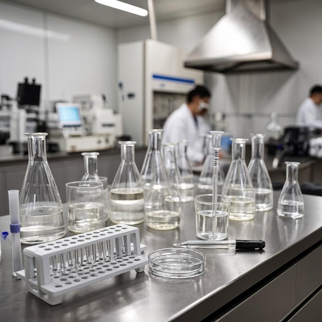
However, for injectable “phase transition” materials that are designed to deliver cells to a wound site, additional and very important, practical criteria exist, which are challenging to meet. First, the gelation kinetics must be fast enough to ensure that cells become homogeneously incorporated within the matrix; it is becoming increasingly clear that cell density plays a role in modulating the behavior of delivered cells. Gelating systems that afford an even distribution of encapsulated cells allow reproducible control over cell density within the matrix. In contrast, systems that gel slowly result in cell sedimentation and gross variation of the cell density throughout the matrix. Second, the spatial resolution with which a gel–cell construct can be introduced in vivo and its ability to remain localized at the point of introduction is of paramount importance. A possible limitation exists for material systems that are delivered to tissue defects as liquids; unless a well defined cavity exists that will contain the hydrogel precursor solution, leakage into/onto neighboring tissue is unavoidable and potentially harmful. For bone and cartilage repair, the implant site can be constrained to limit motion and periosteal flaps can be used to help spatially restrict material leakage. However, even for well defined osteochondral defects, multiple applications of the liquid precursor may be necessary. For other tissues, well defined cavities are not common. In sum, if widespread clinical use is anticipated, then an injectable gel–cell construct must be easily administered, must evenly distribute the delivered cells, and must stay localized at the site of introduction.
We have been developing a general hydrogelation strategy, based on the triggered self-assembly of peptides. The design of this system links the intramolecular folding of amphiphilic β-hairpin peptides to their propensity to self-assemble affording hydrogel material. Peptides are designed such that when dissolved in aqueous solutions, they exist in an ensemble of random coil conformations rendering them fully soluble. However, the addition of an exogenous stimulus results in peptide folding into a β-hairpin conformation that undergoes rapid self-assembly forming a β-sheet-rich, highly cross-linked hydrogel.
Because of electrostatic repulsion between positively charged lysine residues, these peptides remain unfolded in low ionic strength buffer at pH 7.4. However, folding can be triggered by screening some of the lysine-based charge with the addition of DMEM, which contains sufficient concentrations (≈160 mM) of mono- and divalent inorganic salts to ensure effective screening. In the folded state, these peptides adopt a hairpin conformation composed of two β-strand sequences of alternating hydrophobic and hydrophilic residues (Lys and Val) flanking a tetrapeptide type II′ β-turn. These hairpins are amphiphilic molecules in which one face is hydrophobic and the other is hydrophilic. Folded hairpins self-assemble both laterally (via the formation of intermolecular H-bonds and van der Waals contacts) and facially (via the burial of the hydrophobic face of distinct hairpins). Detailed structural characterization indicates that MAX1 gels are comprised of a network of fibrils rich in β-sheet. Each fibril is ≈3 nm in width, consistent with the folded state of the molecule. Fibrils are physically cross-linked by noncovalent, hydrophobic interactions between the hydrophobic faces of assembled hairpins and local fibril entanglements. The fibril persistence length (distance between cross-link sites) ranges from ≈10 to 200 nm. Cryo-transmission electron microscopy (cryo-TEM) and laser scanning confocal microscopy (LCSM) indicate that the gels are well hydrated on both the nano- and microlength scales and are microporous. Together, these material characteristics are attractive for tissue engineering/regeneration applications.
A unique feature of the hairpin gels is that when an appropriate shear stress is applied, the gel will shear-thin, resulting in a low-viscosity gel. However, after the application of shear has stopped, the gel quickly recovers its mechanical rigidity. Shear thin-recovery processes hold promise for minimally invasive material delivery. For example, alginate-based gels impregnated with fibroblast have been delivered s.c. into the backs of rats. However, the measured mechanical rigidity of these gel/cell constructs after syringe delivery indicates that the delivery process is severely detrimental to the mechanical integrity of the gel. In fact, this report concludes that cells may best be delivered as suspensions in alginate solutions that are gelled after syringe delivery.
Here, we report that hairpin hydrogelation can be triggered in the presence of C3H10t1/2 mesenchymal stem cells resulting in self-supporting, mechanically rigid gels that are impregnated with cells. This gel-forming process can be simply performed in a syringe. The resulting gel/cell constructs can then be shear-thin delivered to a targeted secondary site where they quickly recover to their original mechanical rigidity